About This Project
XRF images have high spectral resolution but low spatial resolution, whereas the opposite is true for conventional RGB images. The LR XRF image and the HR RGB image are fused to obtain an HR XRF image.
Project Description
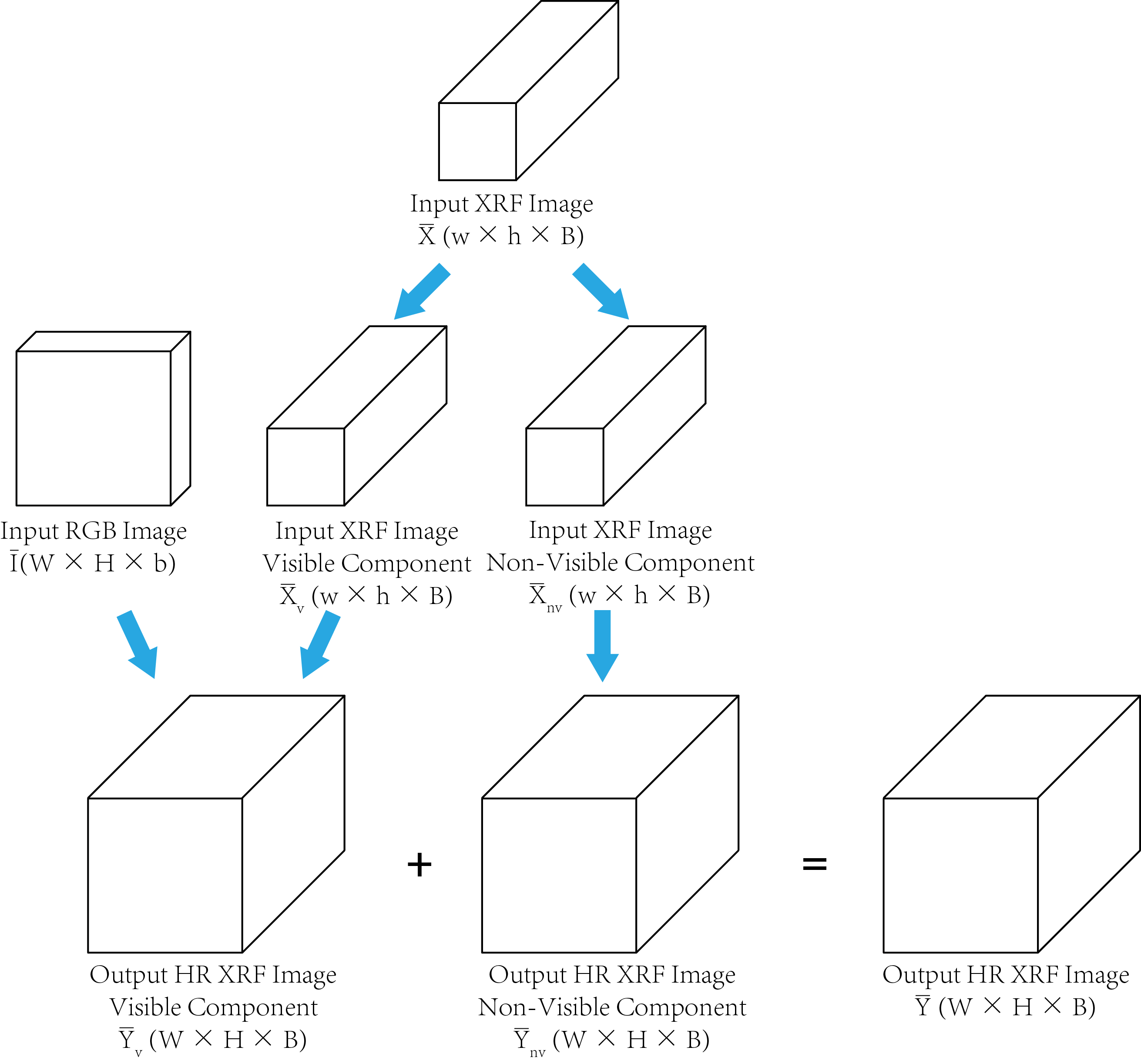
X-Ray fluorescence (XRF) scanning of works of art is becoming an increasingly popular non-destructive analytical method. The high quality XRF spectra is necessary to obtain significant information on both major and minor elements used for characterization and provenance analysis. However, there is a trade-off between the spatial resolution of an XRF scan and the Signal-to-Noise Ratio (SNR) of each pixel’s spectrum, due to the limited scanning time. In this project, we propose an XRF image super-resolution method to address this trade-off, thus obtaining a high spatial resolution XRF scan with high SNR. We fuse a low resolution XRF image and a conventional RGB high resolution image into a product of both high spatial and high spectral resolution XRF image. There is no guarantee of a one to one mapping between XRF spectrum and RGB color since, for instance, paintings with hidden layers cannot be detected invisible but can in X-ray wavelengths. We separate the XRF image into the visible and non-visible components. The spatial resolution of the visible component is increased utilizing the high-resolution RGB image while the spatial resolution of the non-visible component is increased using a total variation superresolution method. Finally, the visible and non-visible components are combined to obtain the final result.
Proposed pipeline of spatial-spectral representation for XRF image super-resolution

The visible component of input XRF image is fused with the input RGB image to obtain the visible component of HR XRF image. The non-visible component of the input XRF image is super-resolved to obtain the non-visible component of HR XRF image. The HR visible and non-visible component of output XRF image are combined to obtain the final
output.
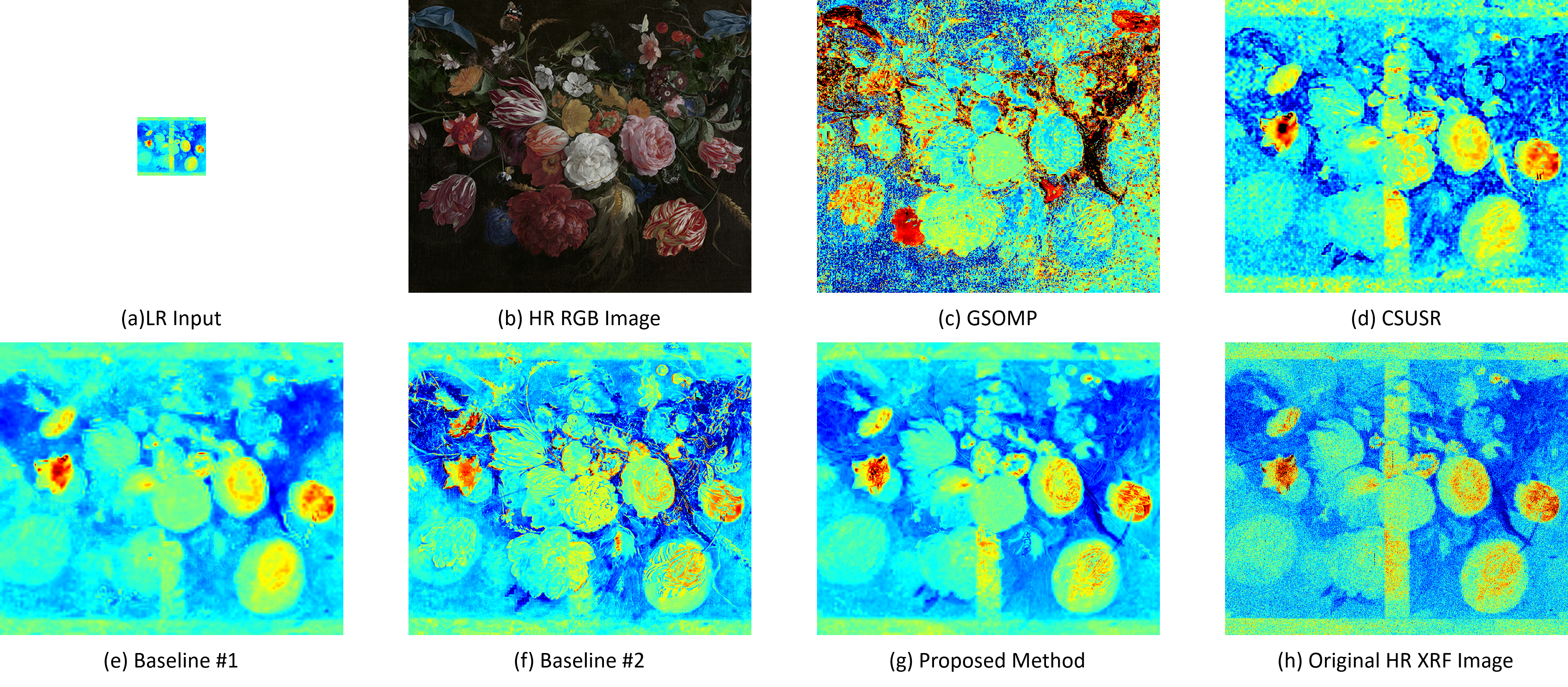
Visualization of the SR result of the DeHeem experiment on the "Bloemen en insecten"

Region of interest of related to Pb Ln XRF emission line (channel #582-602) is selected. (a) is the LR input XRF image and (b) is the HR input RGB image. (c), (d), (e), (f), (g) are the SR result of GSOMP, CSUSR, Baseline #1, Baseline #2 and the proposed method, respectively. (h) is the original HR XRF image.
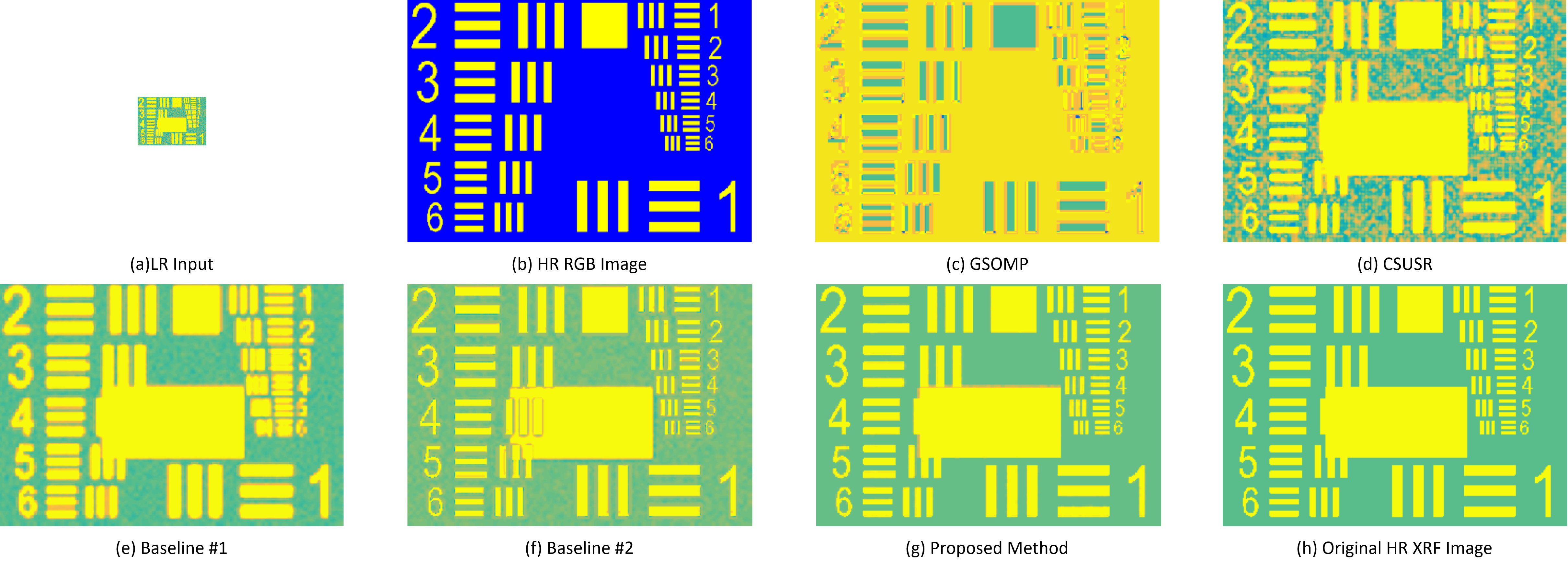
Visualization of the SR result of the Air force synthetic experiment

Visualization of the SR result of the synthetic experiment. Region of interest of channel #210 - 230 is selected. (a) is the LR input XRF image. (b) is the HR input RGB image. (c), (d), (e), (f), (g) are the SR result of GSOMP, CSUSR, Baseline #1, Baseline #2 and the proposed method, respectively. (h) is the original HR XRF image.
Related Publications
"X-Ray fluorescence image super-resolution using dictionary learning"
Qiqin Dai, Emeline Pouyet, Oliver Cossairt, Marc Walton, Francesca Casadio, Aggelos Katsaggelos
Image, Video, and Multidimensional Signal Processing Workshop (IVMSP), 2016 IEEE 12th
"Spatial-Spectral Representation for X-Ray Fluorescence Image Super-Resolution"
Qiqin Dai, Emeline Pouyet, Oliver Cossairt, Marc Walton, and Aggelos K. Katsaggelos
IEEE Transactions on Computational Imaging, Special Issue on Extreme Imaging (preprint)
Back to top


