This exemplary study integrated curatorial, conservation and conservation science research. Curatorial input on visual sources for the artist accompanied the analytical campaign influencing, directing, and inspiring interpretation of the scientific imaging. Two different groups of scientific experts contributed their expertise, thus pushing the limit of interdisciplinary research in a fully integrated framework.
Background
Picasso was a prolific artist throughout the course of his life. One of his earliest periods of production was the Blue Period (1901–1904), so named for his monochromatic paintings in shades of blue and blue-green. Valued for their homogeneous compositions, unified by tone, the paintings of this period are by no means simple, but rather complicated and multi-layered constructs. La miséreuse accroupie (Crouching Woman), painted by Picasso in 1902, now housed at the Art Gallery of Ontario, is a striking example of such complexity, as has been revealed by the recent scientific studies presented on this website. These data are helping art historians to better understand Picasso’s decision making and creative processes at this early and seminal part of his career. In collaboration with the Art Gallery of Ontario (Canada), and the National Gallery of Art, Washington, the Center for Scientific Study of the Arts has undertaken a technical study of La miséreuse accroupie. Here we present some of our results.
The Origins of This Study
Through close observation of La miséreuse accroupie, conservators discovered distinct textures that did not match the visible composition. The presence of an underlying painting was soon proposed as was inspired by the fact that several paintings from the Blue period are known to have been painted on reused supports. This suggestion of another composition beneath La miséreuse accroupie is what inspired the technical study of this painting.
Scientific Tools
X-Ray Radiography
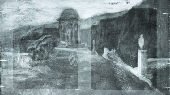
In the 1990’s an x-ray radiograph taken at the Art Gallery of Ontario revealed a hidden landscape painting currently an unknown artist who was painting in Barcelona around the turn of the century. The radiograph revealed this hidden horizontal landscape when the image of the woman is rotated counter-clockwise ninety degrees. It is of note that Picasso incorporated the extant landscape into La miséreuse by reusing some of the lines of the hills as the contours of the woman’s back.
Building a Macro XRF Scanning Stage
The above sample stage (left: front view, right: side view) was originally intended for use by hobbyists interested in computerized numerical controlled (CNC) milling and 3D printing and has been adapted to extend the spatial range previously available to us with the portable XRF instrument from 10 cm² to <100 cm². The basic configuration of the stage is sketched above. For more details about the construction, please contact us.
Hyperspectral Imaging
Diffuse hyperspectral infrared reflectography was performed Dr. John Delaney, Senior Imaging Scientist from the National Gallery of Art, to further investigate the results of traditional infrared reflectography and x-ray radiography. Fiber optic reflectance spectroscopy (FORS) was also used at numerous sites to assist with pigment identification. Hyperspectral image data cubes consisting of around 200 images, each with a spectral range from 967 to 1680 nm, were generated. Significant changes in composition were revealed in the painting.
Photometric Stereo
The surface shape of the painting is composed of submillimeter brush marks, craquelure etc. These data provide information on how the painting was painted. One way to document surface shape is to use reflected light captured from numerous angles as shown our video.
Typically this information is presented as a surface normal vector map, calculated via photometric stereo (PS). A surface normal vector indicates the direction a surface is oriented and is comprised of X, Y, Z gradient maps mapped to RGB channels as may be seen in a cropped portion of the painting above. Note that while this surface lacks significant texture, it still contains rich information about the underlying hidden paint.
X-Ray Fluorescence Imaging
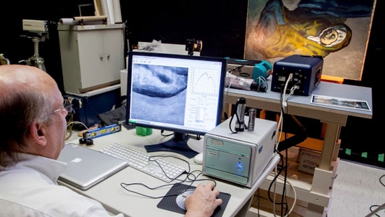
To obtain a more clear understanding of the painting layers, a macro x-ray fluorescence imaging instrument was developed at Northwestern University. To scan large works of art, our team adapted equipment already in use in many cultural heritage institutions, namely a portable XRF instrument, and attached it to an inexpensive in-house built scanning stage with high accuracy. During the experiment, the painting is excited by an x-ray beam approximately 1 mm in diameter. The excitation of the paint generates emission of x-ray fluorescence radiation. The energy of the recorded fluorescence radiation is used to identify the elements present in the analyzed spot. The beam is then moved relative to the painting, in order to scan its surface. The technique allowed us to analyze 70% of the painting in about 24 hours and produce grey scale image maps of the chemical elements characteristic of the pigments present in the different paint layers. The obtained elemental distribution images reveal the spatial positions of various pigments on or beneath the surface of the painting.
Micro-Sampling
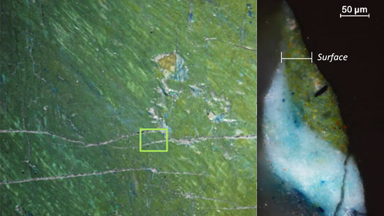
Micro-fragments sampled in strategic places are used to complement the imaging results obtained using non-invasive techniques. The small fragments (of approximatively the size of a hair – a few hundred of microns) are usually embedded into resin and polished to expose the various paint layers. A deeper understanding of the painting stratigraphy is then allowed. Shown above is a cross-section from a fragment sampled from one of the woman’s fingers (pentimentiarea). It clearly illustrates the presence of at least three layers from surface to ground. Upon further microanalysis, the paint composition (pigments, bindersetc.) can be determined.
Complex Structure
Imaging techniques are helping scholars to better understand Picasso’s style, influences and process. Follow the links below to explore the interactive ‘curtain viewers’ which show the overlap of features that become apparent using different modalities of light (from X-rays to visible wavelengths).
Visible miséreuse accroupie
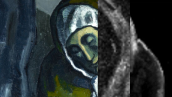
When examining the surface layers of the painting, iron (Fe) and chromium (Cr) based pigments correlate with the painted surface visible to the naked eye. The palette is mostly blues painted with either the iron (Fe) rich Prussian blue or ultramarine. In addition, chrome (Cr) based yellows pigments were used.
View the comparison in the image above: the image on the right is iron (Fe) elemental map, the middle image is chromium (Cr) elemental map, and the image on the left is RGB color.
Earlier Painting
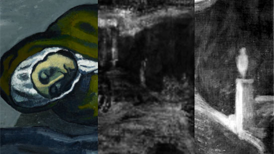
The use of X-rays allows for a deep probing of the paint layers and sheds new light on the composition of the early landscape discovered by radiography. The use of Zinc (Zn), Barium (Ba), Cadmium (Cd), and Mercury (Hg) based-pigments have been identified. Together with micro-analyses of the cross sections, these data will help historians to make comparisons with paintings from the same period and with similar stratigraphic structures.
View the comparison in the image above: the image on the right is X-radiograph, the middle image is zinc (Zn) elemental map, and the image on the left is RGB color.
Pedimenti
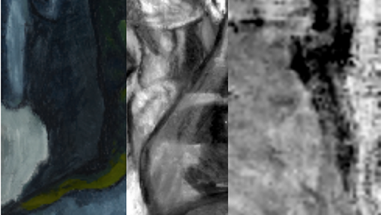
Elemental maps of cadmium (Cd) and lead (Pb) show a slightly altered composition from what is observed at the surface. These pigments reveal a version of the woman with a different head inclination. Together with infrared reflectance hyperspectral images these imaging techniques also revealed the woman’s proper right arm and hand (see the image above). Once you know what you are looking for, these same shapes may be observed in the visible RGB images and radiographs.
After imaging the ‘hand’ area with better spatial resolution using Northwestern’s scanning macro-XRF, it became clear to Kenneth Brummel, the AGO’s assistant curator of modern art, that the hand is similar to that of a crouching woman in a watercolour by Picasso recentlysold at auction (lot 1042). This discovery allowed him to formulate new questions on this intermediate version. The hand itself is also an area that Picasso reworked before abandoning this composition. The iron (Fe) distribution map shows that the third finger of the hand was moved from its original orientation.
Surface Reflectance
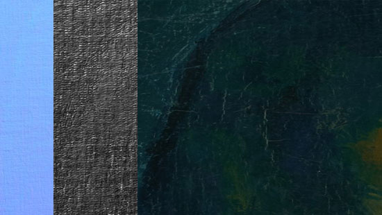
In addition to the color map (albedo) where only diffusely reflected light is recorded, a specularity map (specular albedo or the first bounce of light off the surface) can be calculated from the PS experiment using algorithms developed at Northwestern.
The layered image above shows diffuse color (right), the surface normal vector map (left), or the specular albedo (middle). In these images, one can make out many of the features first recognized in the x-ray and hyperspectral datasets. For instance, in the diffuse albedo image, the intermediate composition made by Picasso one can see the hand grasping the plate. In the specular albedo image, the landscape and temple of the original canvas appear. The specular albedo documents the subtle topographic differences to the top surface imposed by the presence of the underlying composition.
More information about this work and how we captured these data can be found at thecomp photo lab.
Related Talks and Project Media Coverage
Presentation at the American Association for the Advancement of Science 2018 annual meeting: