
About X-ray CT
An X-ray CT scan, or computed tomography scan, is a mature imaging technique to visualize the internal and external three-dimensional volume of an object without physical cutting. The X-ray beam attenuates when passing through the volume of the object. By measuring this attenuation from multiple angles, it reconstructs and renders the cross-sectional (tomographic) images (virtual "slices") to produce the specific volume of a scanned object. A wide range of applications includes biological, anthropological, geological, engineering fields, etc.
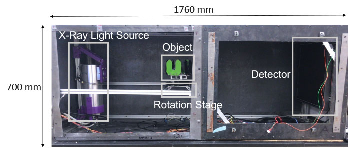
Instrumentation
The CT scanner is built by Tom Bochynek (Mike Rubenstein lab) and Florian Schiffers (Oliver Cossairt lab) and modified by Alexis Baudron (The Computer Science Department, Northwestern University) and Bingjie Xu (NU-ACCESS). It is semi-portable; the X-ray tube allows a maximum voltage of 40 kV and a focal-spot size of 100 or 300 microns.
Applications
X-ray CT can accurately measure the dimension and density of targeted objects. Collaborating with the Avenir Conservation Center at the Denver Museum of Nature and Science (DMNS), we plan to apply CT scanning to assist in establishing the conservation treatments of Conifer Cones, which date back to 130,000 to 70,000 years ago. More details to come soon.
For more information on the Conifer Cones Project at DMNS, please visit:
https://www.dmns.org/science/anthropology/avenir-conservation-center/#ConiferCones